❤️
Cardiac Arrhythmias
Advanced Clinical Lecture
Dr. Shaheena Akram Sheikh
🎓 Final Year Medical Students
Based on Davidson's Principles & Practice of Medicine | ESC Guidelines 2024
🫀Learning Objectives
02 / 17
Learning Objectives
🔬
Understand cardiac electrophysiology and normal conduction system
Explain mechanisms: automaticity, re-entry, triggered activity, conduction block
📊
Identify arrhythmias on 12-lead ECG using a systematic 7-step approach
💊
Describe pharmacological (Vaughan Williams) and interventional treatments (ICD, ablation, PPM)
🫀Introduction
03 / 17
Introduction to Cardiac Arrhythmias

Definition: Any disturbance in rate, rhythm, or conduction of cardiac impulses deviating from normal sinus rhythm (60–100 bpm, regular, P before every QRS).

📊 Epidemiology
  • AF affects ~46 million people worldwide
  • Ventricular arrhythmias → ~300,000 SCDs/year (USA)
  • Leading cardiac mortality cause in patients <40 years
  • Prevalence rises sharply with age
⚠️ Clinical Significance
  • Sudden Cardiac Death – VF/VT most common cause
  • Stroke risk – AF causes 20–30% of ischaemic strokes
  • Heart failure – Tachycardia-induced cardiomyopathy
  • Haemodynamic compromise – Syncope, shock
  • Thromboembolism – AF mural thrombus

⚡ Davidson's Pearl: Haemodynamic stability is the primary determinant of urgency — always assess this before rhythm diagnosis.

Cardiac Electrophysiology
04 / 17
Electrophysiology & Conduction System
⚡ Action Potential Phases
Phase 0 Rapid Na⁺ influx → fast depolarisation
Phase 1 K⁺ efflux → early repolarisation
Phase 2 Ca²⁺ influx + K⁺ efflux → plateau (contraction)
Phase 3 K⁺ efflux → rapid repolarisation
Phase 4 Resting / pacemaker slow depolarisation (If)
🔑 Refractory Periods
  • ARP: No stimulus excites (Phase 0–mid 3)
  • RRP: Strong stimulus triggers AP (late Phase 3)
  • R-on-T: Stimulus during RRP → VF risk ⚠️

SA Node: 60–100 bpm via If. AV Node: delays 120ms. Bundle of His → L/R bundles → Purkinje (20–40 bpm escape).

RA LA RV LV SA Node 60–100 bpm AV Node 40–60 bpm · 120ms delay Bundle of His RBB LBB Purkinje Fibres · 20–40 bpm
📊Normal ECG & Interpretation
05 / 17
Normal ECG – Waveforms, Intervals & 7-Step Method
Lead II – Normal Sinus Rhythm (Rate 72 bpm)
P QRS T ST segment PR 120–200ms QRS <120ms QTc <440ms ♂ / <460ms ♀
🔢 7-Step Method
1
Rate – 300/large squares (regular). ×6 in 10s (irregular)
2
Rhythm – Regular? Regularly vs irregularly irregular?
3
P waves – Present, normal, 1:1 with QRS, upright in I & II?
4
PR interval – 120–200ms. Short=WPW. Long=AV block
5
QRS – <120ms. Wide=BBB or VT. LBBB vs RBBB morphology
6
QTc – Bazett QT/√RR. >500ms = Torsades risk
7
Axis – Normal –30° to +90°. NW axis → VT

⚡ Golden Rule: Wide-complex tachycardia (QRS >120ms, rate >100) = VT until proven otherwise. Apply Brugada criteria.

Wave Summary
  • P wave – Atrial depolarisation. <120ms, <2.5mm
  • QRS – Ventricular depolarisation. Pathological Q >1mm wide
  • ST segment – Isoelectric. Elevation=STEMI, depression=ischaemia
  • T wave – Ventricular repolarisation. Inversion=ischaemia/BBB
♾️Mechanisms of Arrhythmia
06 / 17
Pathophysiological Mechanisms
🔄 1. Abnormal Automaticity
  • Enhanced: Ischaemia, catecholamines, hypokalaemia → ectopic foci fire faster than SA node
  • Suppressed: Vagal tone, drugs → sick sinus syndrome, bradycardia
  • Abnormal Phase 4 slope in non-pacemaker cells
⚡ 2. Triggered Activity
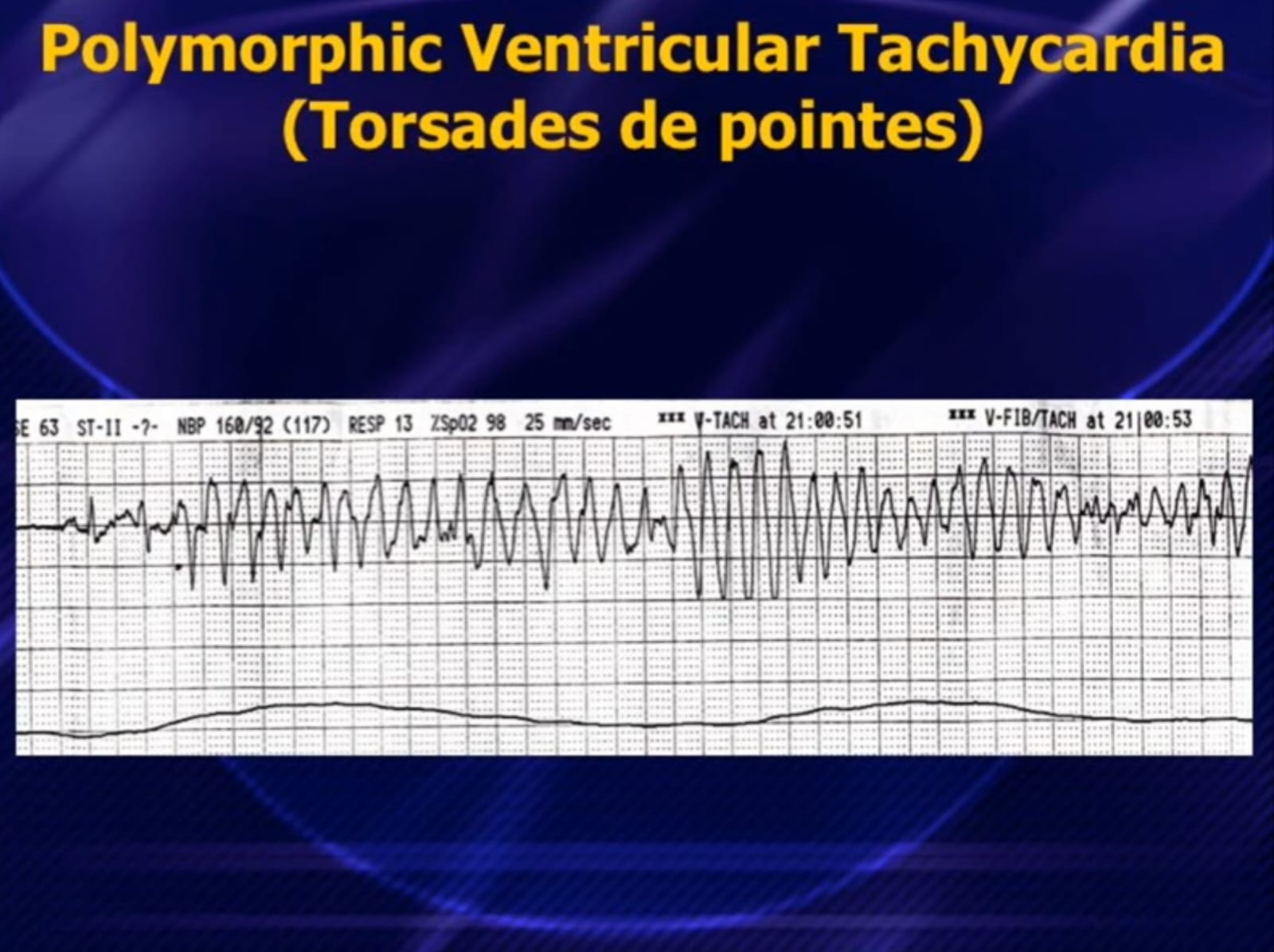
  • EADs: Phase 2–3. Long QT, drugs, hypokalaemia → Torsades de Pointes
  • DADs: Phase 4. Ca²⁺ overload → Digitalis toxicity, catecholamines
♾️ 3. Re-entry Circuit ← Most Common!
1
Two pathways with different refractory periods
2
Unidirectional block in one pathway
3
Slow conduction → recovery of blocked path → circus movement
Examples: AF, Atrial Flutter, AVNRT, AVRT, Scar-VT
🚧 4. Conduction Block
  • Failure of impulse transmission: fibrosis, ischaemia, drugs, vagal tone
  • Results: AV blocks (1°/2°/3°), BBB, SA exit block

Davidson's: Re-entry is the mechanism of most sustained arrhythmias. Catheter ablation targets and interrupts the circuit — basis of curative ablation.

📋Classification of Arrhythmias
08 / 44
Classification of Cardiac Arrhythmias
🔵 Supraventricular (SVA)
Tachy
  • Sinus Tachycardia
  • Atrial Fibrillation (AF)
  • Atrial Flutter
  • Atrial Tachycardia
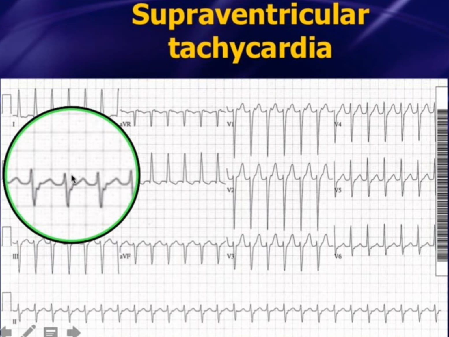
  • AVNRT (most common SVT)
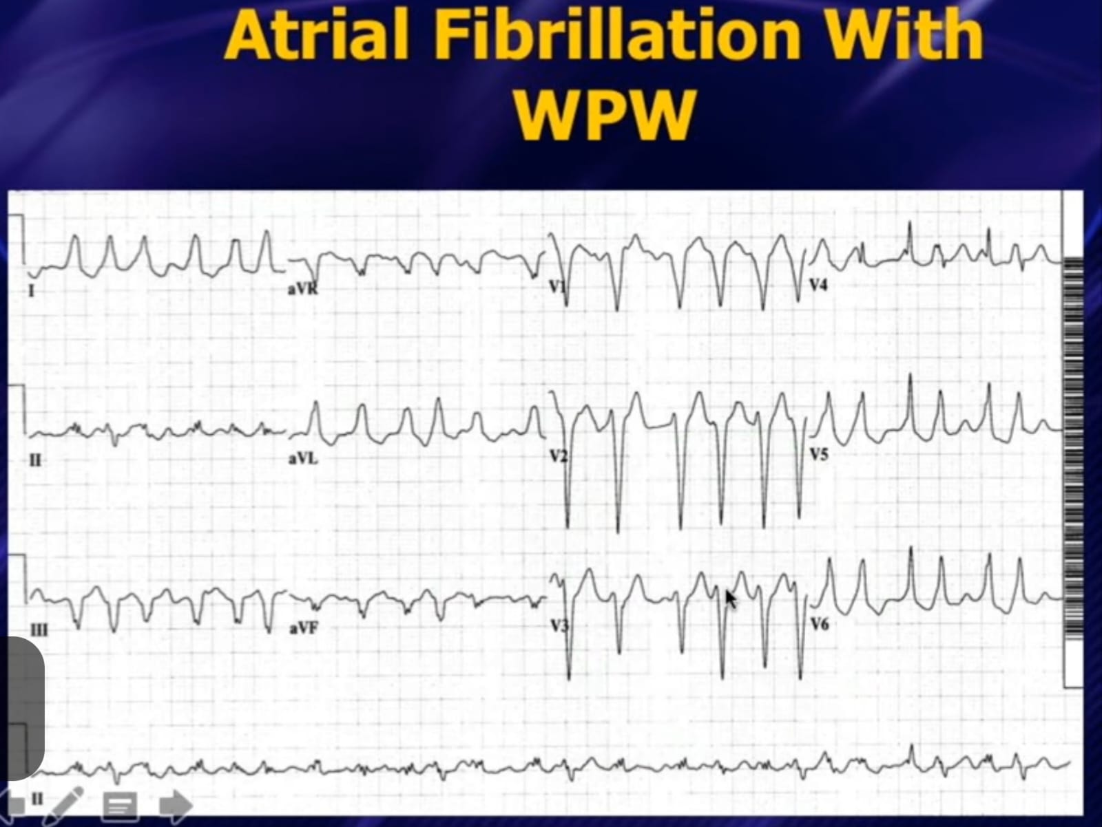
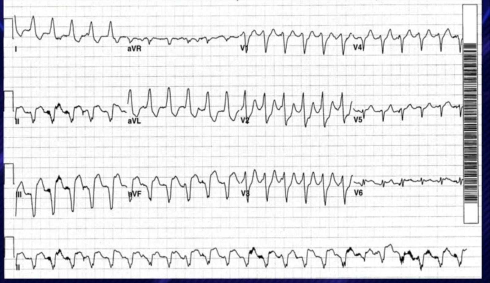
  • AVRT (WPW syndrome)
  • Junctional Tachycardia
Brady
  • Sinus Bradycardia
  • Sick Sinus Syndrome
🔴 Ventricular (VA)
Tachy
  • Premature Ventricular Complexes (PVCs)
  • Ventricular Tachycardia (VT)
  • Sustained vs Non-sustained VT
  • Monomorphic vs Polymorphic VT
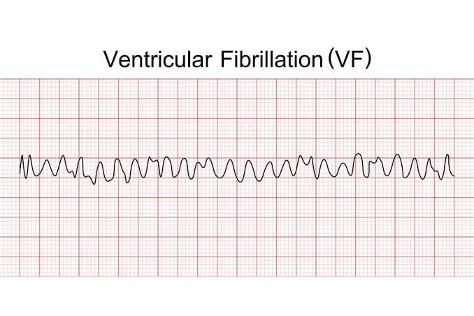
  • Ventricular Fibrillation (VF)
  • Torsades de Pointes
  • Idioventricular Rhythm
🟡 Conduction Disorders
AV Blocks
  • 1st Degree AV Block
    PR >200ms, all conducted
  • 2nd Degree – Mobitz I
    Wenckebach – progressive PR ↑ then dropped beat
  • 2nd Degree – Mobitz II
    Sudden non-conducted P wave – dangerous!
  • 3rd Degree (Complete)
    Complete dissociation – P & QRS independent
BBB
  • RBBB / LBBB / Fascicular Blocks
📊ECG – Atrial Fibrillation & Flutter
07 / 17
Atrial Fibrillation & Atrial Flutter
Lead II – Atrial Fibrillation (Irregularly Irregular · No P Waves · f waves)
✕ No P Waves Fibrillatory baseline (f waves 350–600/min) Irregularly Irregular RR intervals — hallmark of AF
Lead II – Atrial Flutter · 4:1 Block · Atrial rate ~300 bpm · Ventricular ~75 bpm
Classic "Sawtooth" Flutter Waves — Re-entry in cavotricuspid isthmus Regular QRS every 4th flutter wave · 2:1 most common → ventricular rate ~150 bpm 4 F-waves
AF Management
  • CHA₂DS₂-VASc ≥2(♂)/≥3(♀) → DOAC anticoagulation
  • Rate control: β-blocker/diltiazem/digoxin <110 bpm
  • Rhythm control: DCCV, flecainide (no structural disease), amiodarone
Flutter Management
  • Cavotricuspid isthmus ablation — >95% cure rate
  • Rate/rhythm control as per AF guidelines
  • Anticoagulate by CHA₂DS₂-VASc score
📊ECG – Atrial Fibrillation & Flutter (Examples)
08 / 18
12-Lead ECG Examples
12-lead ECG – Atrial Fibrillation
12-Lead ECG · Atrial Fibrillation (irregularly irregular, no P waves, fibrillatory baseline)
ECG strip – Atrial Flutter
Rhythm strip · Atrial Flutter (sawtooth F waves, regular QRS)
📊ECG – SVT & WPW Syndrome
09 / 18
SVT (AVNRT) & WPW Syndrome
Lead II – SVT/AVNRT · Rate ~180 bpm · Regular · Narrow Complex · No visible P waves
Regular narrow QRS · P waves hidden IN QRS (retrograde) · Abrupt onset/offset Mechanism: Dual AV nodal pathway re-entry (AVNRT) · Treatment: Vagal → Adenosine 6mg IV → Ablation
Lead I – WPW Pattern · Short PR · Delta (δ) Wave · Wide QRS · Pre-excitation
δ δ δ Bundle of Kent bypasses AV node → Short PR + Delta wave + Wide QRS = Pre-excitation ⚠️ AF+WPW = DANGER → avoid adenosine/verapamil/digoxin!
SVT Management
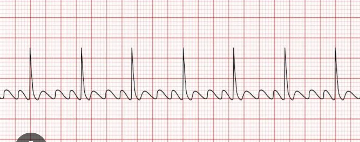
  • Vagal manoeuvres → IV Adenosine 6mg bolus (terminates >90%)
  • Alternative: IV Verapamil / β-blocker
  • Unstable: Synchronised DCCV | Curative: RF ablation >98%
⚠️ WPW Critical Points
  • AF+WPW: rapid accessory conduction → VF risk (200–300 bpm)
  • Treat with IV Procainamide or DC cardioversion
  • Definitive: Pathway ablation — curative
📊ECG – SVT & WPW (Examples)
10 / 21
12-Lead ECG Examples – SVT & WPW
12-lead ECG – Supraventricular tachycardia
Supraventricular tachycardia · Narrow complex, regular rhythm
12-lead ECG – Atrial Fibrillation with WPW
Atrial Fibrillation with WPW · Pre-excitation, irregular wide QRS
🚨ECG – VT & Ventricular Fibrillation
09 / 18
Ventricular Tachycardia & Ventricular Fibrillation
Lead V1 – Monomorphic VT · Rate ~150 bpm · Wide QRS >160ms · AV Dissociation · LIFE-THREATENING
WIDE QRS >160ms · Bizarre Morphology · Extreme Axis · Concordance in V leads ↑ Independent P waves (AV dissociation) = diagnostic of VT
Ventricular Fibrillation — CARDIAC ARREST — No QRS — CPR + Defibrillation IMMEDIATELY
Chaotic disorganised waveforms — No identifiable QRS — No cardiac output
🚨 VT Management
  • Unstable: Synchronised DCCV 200J biphasic (sedate first)
  • Pulseless VT: CPR + unsynchronised defibrillation
  • Stable VT: IV Amiodarone 300mg over 20–60 min
  • Correct K⁺ >4.5, Mg²⁺ >1.0 | Long-term: ICD if EF <35%
🚨 VF – ACLS
  • CPR immediately (100–120/min, 5–6cm)
  • Unsynchronised 360J mono / 200J biphasic
  • Adrenaline 1mg IV every 3–5 min after shock 3
  • Amiodarone 300mg IV after shock 3
  • 4Hs & 4Ts reversible causes
🚨ECG – VT & VF (Examples)
12 / 21
12-Lead ECG Examples – VT & Ventricular Fibrillation
12-lead ECG – Ventricular Tachycardia
Ventricular Tachycardia · Wide QRS, regular monomorphic
ECG – Ventricular Fibrillation
Ventricular Fibrillation · Chaotic, no identifiable QRS
📊ECG – Torsades de Pointes & Long QT
10 / 17
Torsades de Pointes & Long QT Syndrome
Lead II – Long QT → R-on-T → Torsades de Pointes (Polymorphic VT twisting around isoelectric line)
QTc >500ms = HIGH RISK R-on-T → Twisting QRS amplitude — "turning of the points" — may degenerate to VF
Long QT Causes
  • Congenital: LQT1 (KCNQ1), LQT2 (KCNH2), LQT3 (SCN5A)
  • Drugs: Amiodarone, sotalol, quinidine, macrolides, antipsychotics
  • Electrolytes: ↓K⁺, ↓Mg²⁺, ↓Ca²⁺
  • Other: Bradycardia, hypothyroidism, intracranial events
✅ Torsades Management
  • First-line: IV MgSO₄ 2g over 10 min (even if Mg²⁺ normal)
  • Stop all QT-prolonging drugs immediately
  • Correct K⁺ >4.5 mmol/L
  • Overdrive pacing 100 bpm if recurrent
  • Congenital LQTS: β-blockers (LQT1/2), ICD (high risk)

QTc >500ms = very high risk for TdP. Always check drug chart!

📊ECG – Torsades de Pointes (Example)
14 / 21
Polymorphic VT – Torsades de Pointes
ECG – Torsades de Pointes
Torsades de Pointes · Polymorphic VT, twisting QRS around baseline
📊ECG – AV Blocks
11 / 17
Atrioventricular (AV) Blocks
1° AV Block — Long PR (>200ms) · All P waves conducted · Benign
PR >200ms Every P followed by QRS · No treatment usually needed
2° Mobitz I (Wenckebach) — Progressive PR ↑ → Dropped beat · Grouped beating
P–no QRS Progressive PR lengthening → dropped QRS → grouped beating
2° Mobitz II — Sudden Dropped QRS without warning ⚠️ Risk of complete block!
P, no QRS! P, no QRS! P, no QRS! Constant PR · Sudden block · Pacemaker needed!
3° Complete AV Block — P waves & QRS completely independent · PACEMAKER mandatory
P waves (fast · regular) Slow escape rhythm (wide · independent) No relationship between P & QRS

Management: 1° → observe | Mobitz I → monitor/treat cause | Mobitz II & 3° → IV Atropine bridge → Permanent Pacemaker (Class I indication)

🏥Clinical Presentation & Diagnostics
12 / 17
Clinical Presentation & Diagnostic Approach
💓 Symptoms
  • Palpitations – regular fast (SVT), irregular (AF), missed beats (ectopics)
  • Syncope – Stokes-Adams (complete block), exertional (VT/HCM) = RED FLAG
  • Pre-syncope/Dizziness – reduced cardiac output
  • Chest pain – tachycardia-induced ischaemia
  • Dyspnoea – haemodynamic compromise, decompensated HF
  • Asymptomatic – incidental (PVCs, 1° AVB, paroxysmal AF)

⚡ Red Flags: Exertional syncope · Family history SCD · Structural heart disease · Syncope without prodrome · Wide complex tachycardia

1
12-Lead ECG – First-line. Pre-excitation, QT, LVH, Q waves, BBB
2
24h Holter – Daily symptoms. PVC burden, silent AF
3
Event/Loop Recorder – ILR (3yr) = gold standard unexplained syncope
4
Echo – EF (ICD threshold), structural disease. Mandatory pre-DCCV
5
EPS – Invasive mapping + ablation. SVT, VT, WPW risk stratification
6
Bloods – TFTs, K⁺, Mg²⁺, Ca²⁺, troponin, digoxin level

Always correct: K⁺, Mg²⁺, thyroid, drugs, ischaemia before escalating treatment.

🏥Interactive Clinical Case
15 / 17
Clinical Case – What is the Diagnosis?
Presentation
68M · Hypertension · T2DM · Regular alcohol. 4h palpitations + breathlessness. Haemodynamically stable. BP 135/82 · HR 118 (irregular) · SpO₂ 97%
Bloods
TSH 0.3 (↓) · K⁺ 3.8 · Mg²⁺ 0.8 · Troponin negative · Normal renal function
ECG – What is the rhythm?
No P waves · Irregularly Irregular QRS · Narrow complex
❓ Discussion Questions
Q1
What is the ECG diagnosis? State the 3 criteria.
Q2
What are the precipitants in this patient?
Q3
CHA₂DS₂-VASc score? Anticoagulation needed?
Q4
Immediate management plan?
✅ Answers
  • Diagnosis: New-onset AF — absent P waves, irregular narrow QRS, f-wave baseline
  • Precipitants: Hyperthyroidism (TSH low) + alcohol — both reversible
  • CHA₂DS₂-VASc: Age(1)+HTN(1)+DM(1) = 3 → DOAC strongly indicated (apixaban)
  • Mx: Rate control (bisoprolol 2.5mg), DOAC, treat hyperthyroidism, TTE, consider cardioversion after 3wk anticoagulation
Key Takeaways
16 / 17
Summary – Key Clinical Points
🎯
Haemodynamic stability determines urgency — unstable = immediate electrical therapy regardless of rhythm
📊
Wide complex tachycardia = VT until proven otherwise — apply Brugada criteria
💊
Adenosine is diagnostic AND therapeutic for SVT — NEVER in AF+WPW (precipitates VF)
🔄
Re-entry drives most sustained arrhythmias — catheter ablation interrupts the circuit
🛡️
AF anticoagulation: CHA₂DS₂-VASc ≥2(♂)/≥3(♀) → DOAC; balance with HAS-BLED
Torsades: IV MgSO₄ 2g first-line. Remove QT drugs. QTc >500ms = high risk marker
🔋
ICD prevents SCD in EF ≤35%. Ablation cures SVT/AFL/WPW. PPM mandatory for complete AV block
🧪
Correct reversible causes: K⁺, Mg²⁺, thyroid, drugs, ischaemia — always before escalating therapy
📚References
17 / 17
References & Further Reading
📖 Textbooks
  • Davidson's Principles & Practice of Medicine, 23rd Ed. Elsevier 2022
  • Braunwald's Heart Disease, 12th Ed. Elsevier 2022
  • Kumar & Clark's Clinical Medicine, 10th Ed. 2022
  • Hurst's The Heart, 14th Ed. McGraw-Hill 2022
🏛️ Guidelines
  • ESC AF Guidelines 2020 – Van Gelder et al. Eur Heart J 2020
  • ESC Ventricular Arrhythmia & SCD 2022 – Zeppenfeld et al.
  • ESC Cardiac Pacing 2021 – Glikson et al.
  • AHA/ACC/HRS SVT Guidelines 2015 – Page et al.
  • AHA ACLS 2020 | Resuscitation Council UK 2021
Dr. Shaheena Akram Sheikh
Thank you for your attention | Questions & Discussion Welcome
"The heart has its own electricity – understanding it is the foundation of clinical cardiology"
❤️